
DC-100
A three-layer integrated platform closing the loop from staining to reading.
Models DC120 (high-throughput floor-standing) and DC40 (compact benchtop). Technical principle: liquid-based cytology with natural sedimentation. Applications: early cervical cancer screening (TCT); gynecological and non-gynecological exfoliated cytology (pleural and ascitic fluids, urine, sputum, etc.).
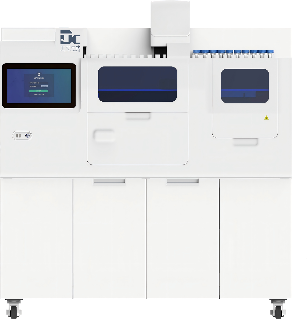
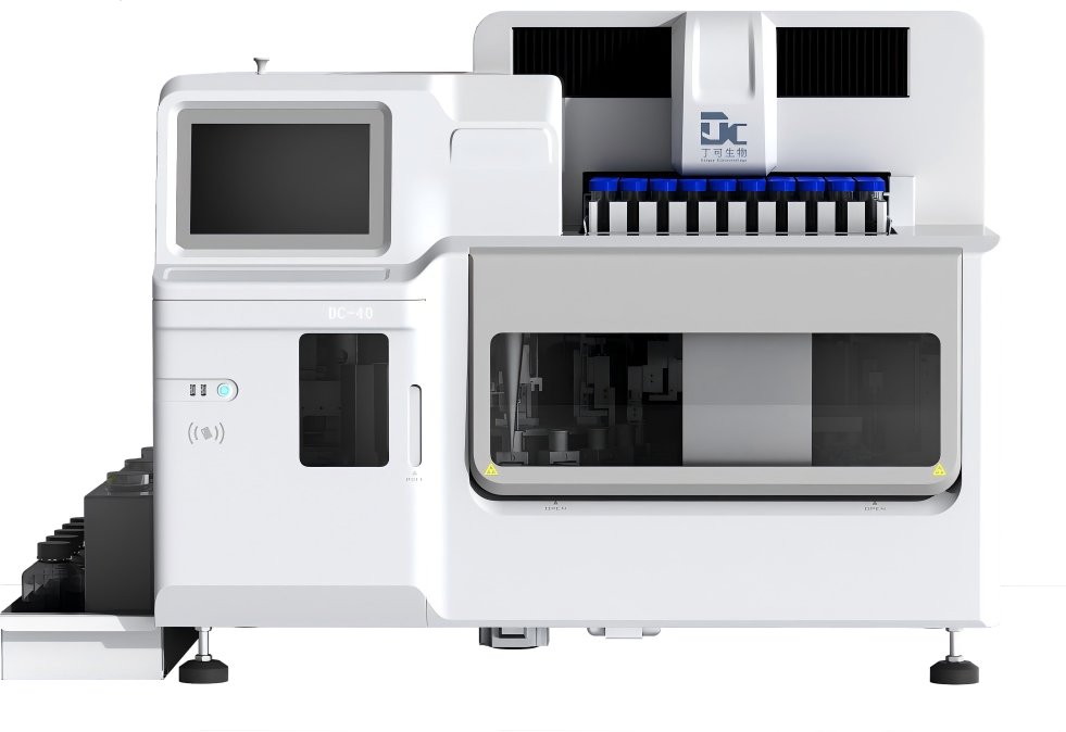
High throughput: up to 120 slides/hour for high-volume laboratories
Ease of use: random-access loading, LIS/HIS connectivity, on-site fault recovery and power-off memory
Reliability: fully automated dripping stain, disposable sedimentation chambers, 100 μl pipetting precision, triple self-cleaning sample probes
Full automation: automatic sample scanning and slide barcoding, automated chamber handling, preparation and staining
| Specification | DC120 | DC40 |
|---|---|---|
| Throughput | 120 slides/hour | 40 slides/hour |
| Samples in parallel | 50 (continuous loading) | 50 |
| Chamber auto-sorting capacity | ≥300 | ≥150 |
| Slide / coverslip loading | ≥100 / ≥250 | ≥100 / ≥250 |
| Slide collection | 120 slides | 90 slides |
| STAT positions | — | 10 |
| Diagnostic area | ~15 mm in diameter | ~15 mm in diameter |
| Auto loading/preparation/staining/coverslipping | √ | √ |
| Auto slide barcoding | √ | √ |
| LIS/HIS connectivity | √ | √ |
| Manual intervention | None | None |
| Form factor | Floor-standing | Benchtop |
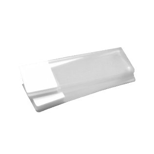
Disposable sedimentation chambers — one chamber per sample; four alternating sampling probes with triple cleaning cycles.
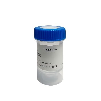
Preserves cell morphology; mucolysis and RBC lysis; long shelf life
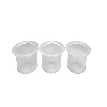
One chamber per sample to prevent cross-contamination
Strong adhesion, minimal cell loss
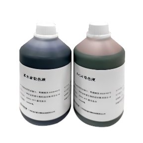
Rich staining with clear N/C contrast and stable performance
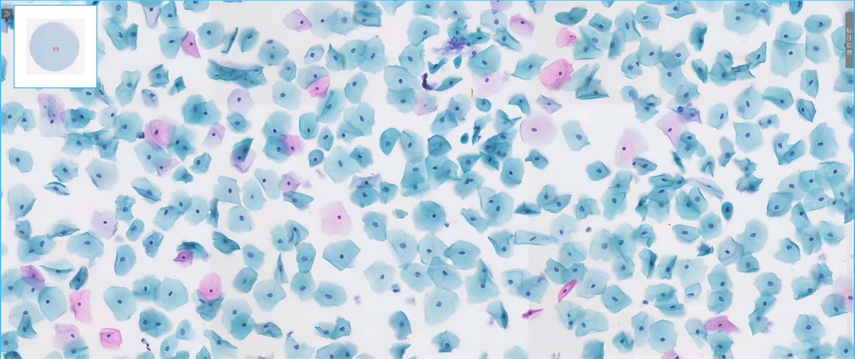
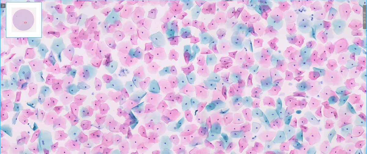
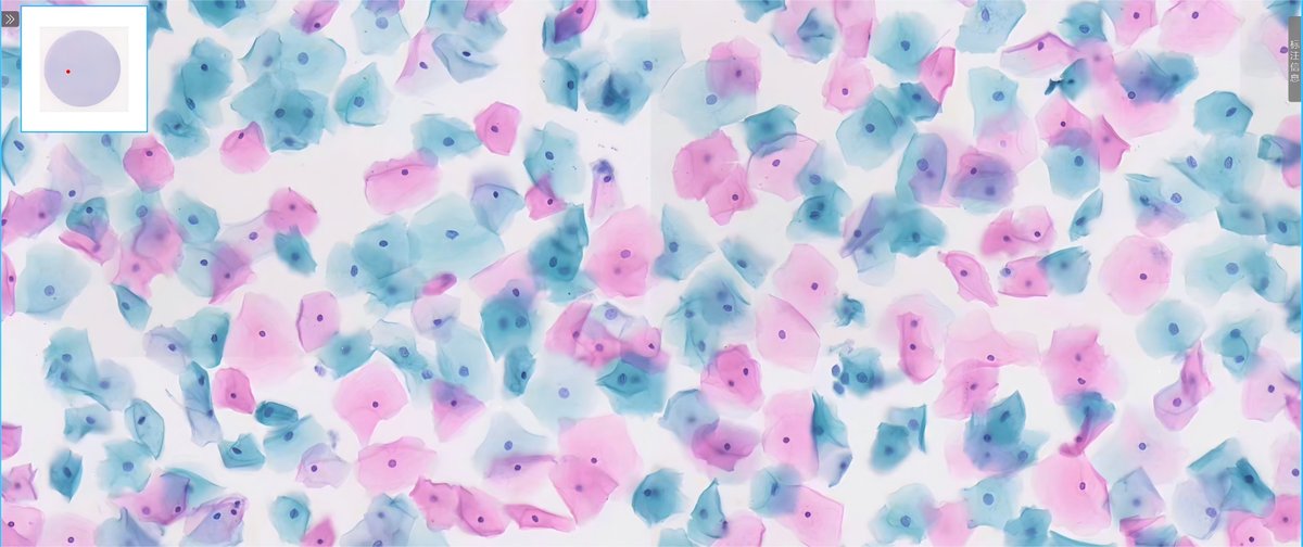
Pap-stained slides show a homogeneous monolayer with clear nuclear-cytoplasmic contrast.
A three-layer integrated platform closing the loop from staining to reading
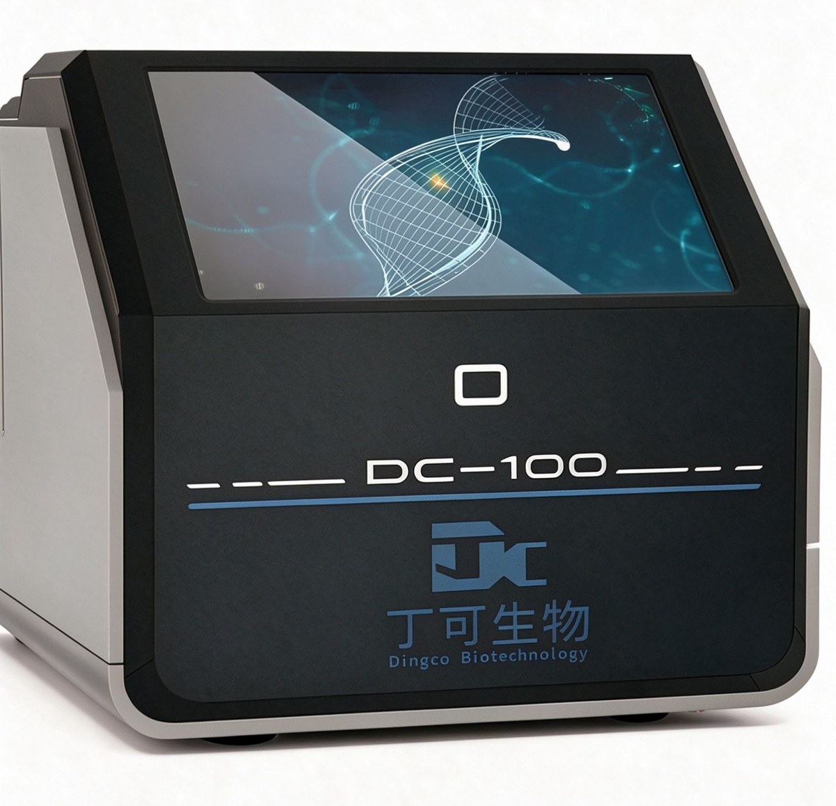
A three-layer integrated platform closing the loop from staining to reading.
Acridine-orange-based one-step staining; dual-color fluorescence (green nuclear DNA / orange-red cytoplasmic RNA); 12-month shelf life, ready to use
Whole-slide scanning with a 20× plan-achromatic objective; barcode/QR recognition with LIS connectivity; auto-focus, image stitching and threshold segmentation
CNN-based deep learning trained on millions of annotated images; multi-dimensional morphology/color/brightness recognition; AI pre-screening → pathologist review → cloud-based quality control
Fluorescent staining of vaginal discharge for BV/AV/VVC/TV and mixed infections, Nugent scoring
Kappa 0.896 vs. Gram stain (n=265)
Non-invasive detection of urine exfoliated cells for bladder cancer screening and follow-up
Sensitivity 77%–85.5% (published literature)
Fluorescence-assisted diagnosis of fine-needle aspirates, report in ~10 minutes in the aspiration room
Sensitivity/specificity 96.3%/93.8% (single-center internal data, external validation pending)
Fluorescent staining for cutaneous/pulmonary fungi and BALF samples
Recommended by ESCMID-ECMM-ERS 2017 guideline